
Aliviar Mezquita Arne abdominal anatomy diagram curva Nuevo significado
The vulva is the global term that describes all of the structures that make the female external genitalia. The components of the vulva are the mons pubis, labia majora, labia minora, clitoris, vestibular bulbs, vulva vestibule, Bartholin's glands, Skene's glands, urethra, and vaginal opening. The mons pubis is a tissue mound made up of fat.

Human Female Anatomy Diagram Human Female External Anatomy Bodemawasuma
Browse 2,691 female stomach anatomy photos and images available, or start a new search to explore more photos and images. Browse Getty Images' premium collection of high-quality, authentic Female Stomach Anatomy stock photos, royalty-free images, and pictures. Female Stomach Anatomy stock photos are available in a variety of sizes and formats.

Pin on Body & Soul
ID: exh6130a. This medical illustration depicts a mid-sagittal view of the normal anatomy of the female abdomen and pelvis. Labeled structures include the large bowel (colon or large intestine), umbilicus, small intestine, ovary, fallopian tube, uterus and bladder.
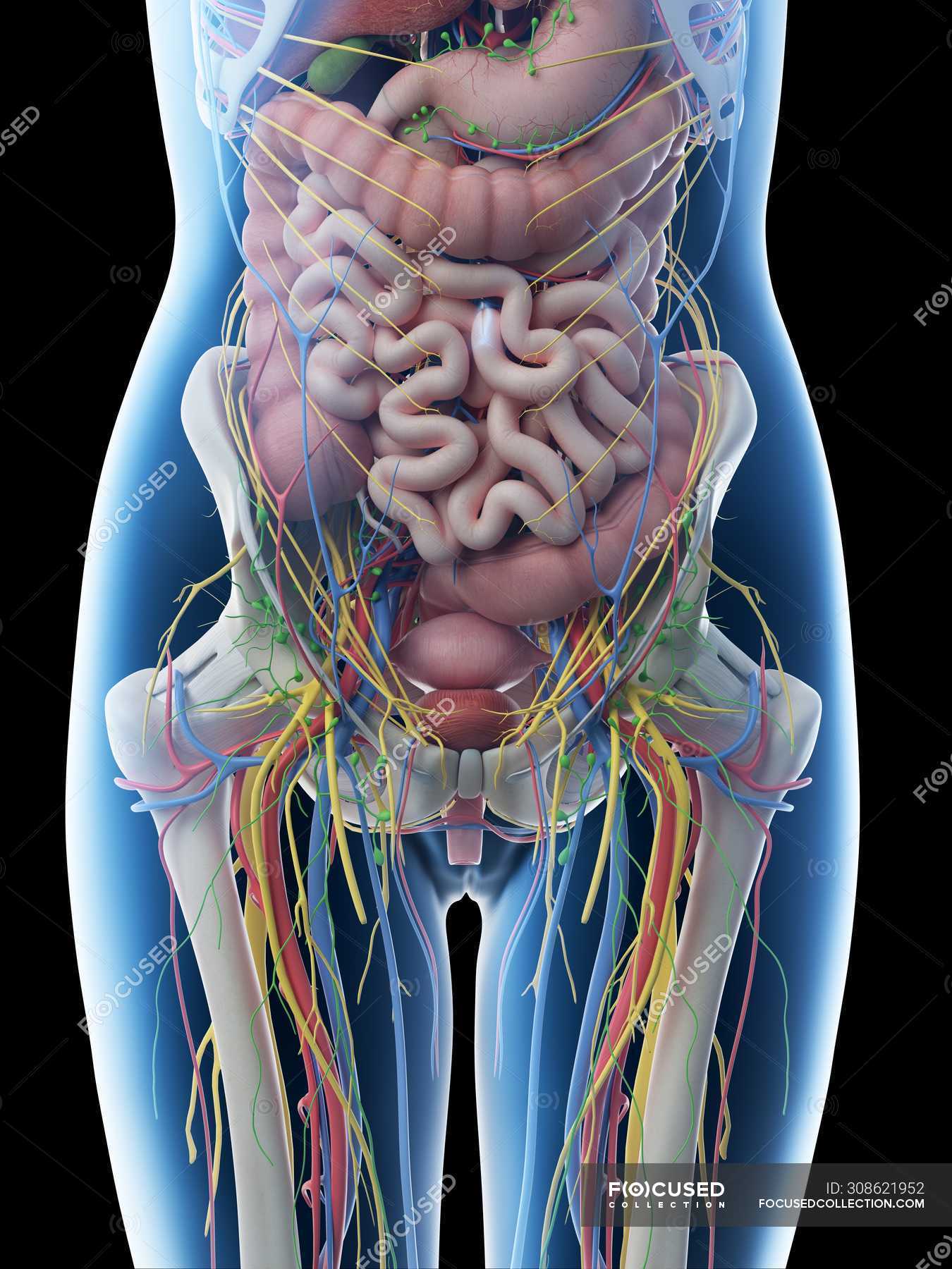
Female abdominal anatomy and internal organs, computer illustration
Anatomy of Female Pelvic Area. Endometrium. The lining of the uterus. Uterus. Also called the womb, the uterus is a hollow, pear-shaped organ located in a woman's lower abdomen, between the bladder and the rectum. Ovaries. Two female reproductive organs located in the pelvis. Fallopian tubes.

Anatomia Kobiety Jelita Zdjęcia ze zbiorów
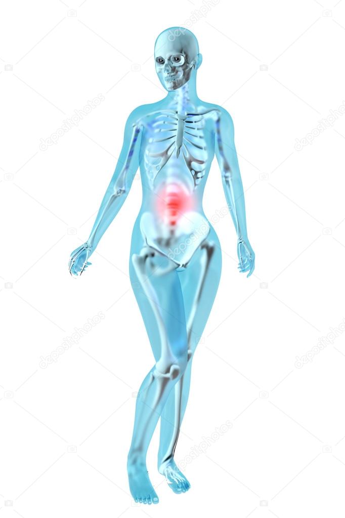
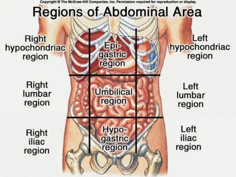
Picture of Abdomen. The abdominal cavity is the part of the body that houses the stomach, liver, pancreas, kidneys, gallbladder, spleen, and the large and small intestines. The diaphragm marks the top of the abdomen and the horizontal line at the level of the top of the pelvis marks the bottom. Connective tissue called the mesentery holds the.

The Anatomy of the Abdomen Human Stomach Health Life Media
Breasts. Summary. Female anatomy includes the external genitals, or the vulva, and the internal reproductive organs, which include the ovaries and the uterus. One major difference between males.
Anatomy Of Stomach Female
Weeks 9 to 12. Where it really starts to get interesting is around weeks 9 through 12. If you even have a baby bump, it'll probably be teeny because your uterus is smushing your intestines together to make room. Fast forward to week 40, and those same intestines take up just a fraction of the space — that's why it's so hard to eat more than a.
Anatomy Of Stomach Anatomy Book
The muscles of the abdomen protect vital organs underneath and provide structure for the spine. These muscles help the body bend at the waist. The major muscles of the abdomen include the rectus.

Real Human Stomach Anatomy Images and Photos finder
The bladder, also known as the urinary bladder, is an expandable, muscular sac that stores urine. When signaled, the bladder releases urine into the urethra, a tube that carries it out of the body.

Stomach Diagram Woman Wiring Diagram
Regardless of their differences, both women and men can improve or maintain the health of their digestive system by maintaining a healthy lifestyle, Dr. Singh says. "Everyone should drink plenty of water, about 64 ounces a day on average, and eat a nutritious diet that includes foods that are high in fiber. The ideal is 25 grams of fiber per day.
Anatomy Of The Female Abdomen And Pelvis, Cut away View Healthiack
The female reproductive system is an intricate arrangement of structures that can separate into external and internal genitalia. The external genitalia comprises the structures outside of the true pelvis, including the labia majora and minora, vestibule, Bartholin glands, Skene glands, clitoris, mons pubis, perineum, urethral meatus, and periurethral area. The internal genitalia is the.
/images/chapter/lymphatics-of-abdomen-and-pelvis/Lymphatics_of_abdomen_and_pelvis_2.png)
Abdominal Anatomy Organs Anatomy Of The Female Abdomen And Pelvis
The pelvic cavity is a bowl-like structure that sits below the abdominal cavity. The true pelvis, or lesser pelvis, lies below the pelvic brim (Figure 1). This landmark begins at the level of the sacral promontory posteriorly and the pubic symphysis anteriorly. The space below contains the bladder, rectum, and part of the descending colon. In females, the pelvis also houses the uterus.
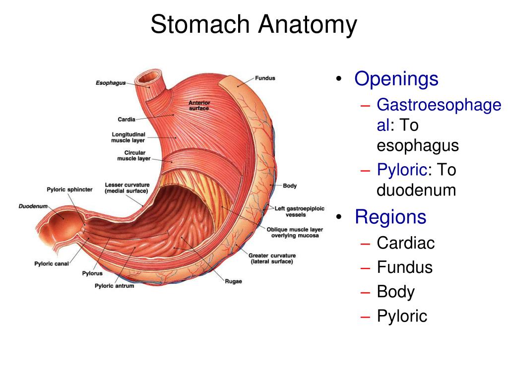
PPT Stomach Anatomy PowerPoint Presentation, free download ID1940155
Matej G. is a health blogger focusing on health, beauty, lifestyle and fitness topics. He has been with healthiack.com since 2012 and has written and reviewed well over 500 coherent articles.
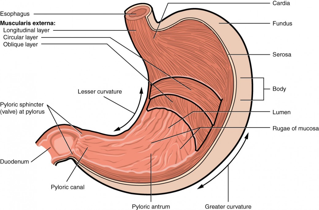
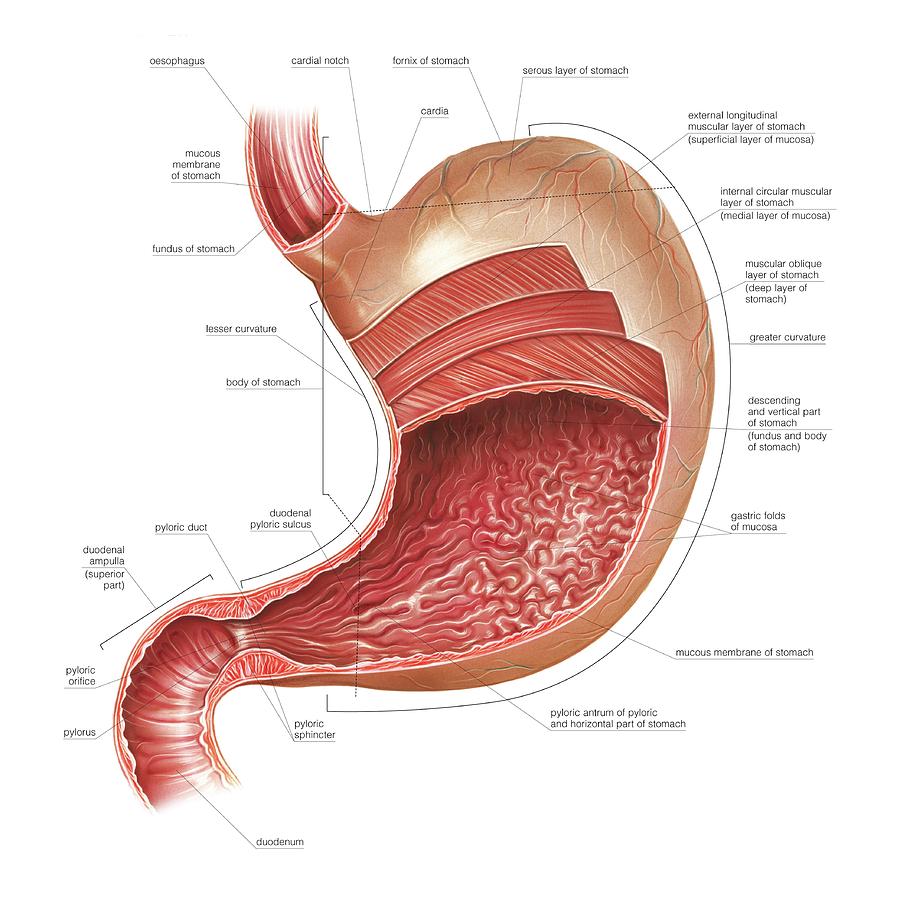
The Stomach Anatomy and Physiology II
Female anatomy includes the internal and external structures of the reproductive and urinary systems. Reproductive anatomy plays a role in sexual pleasure, getting pregnant, and breastfeeding. The urinary system helps rid the body of toxins through urination (peeing). The main parts of the female anatomy can be broken up into outside (external.

Anatomy of female stomach, illustration Stock Image F010/9291
Stomach. The stomach is on the upper-left area of the abdomen below the liver and next to the spleen. It stores and breaks down the foods and liquids we eat before they move to digestion. When the.

Stomach Anatomy Medical Art Library
The stomach is located in the upper part of the abdomen. The digestive organs in the abdomen work together to absorb nutrients and move food through the digestion process. They include the stomach.