PPT The Nose and Para nasal Sinuses 241 RTS PowerPoint Presentation, free download ID1484813
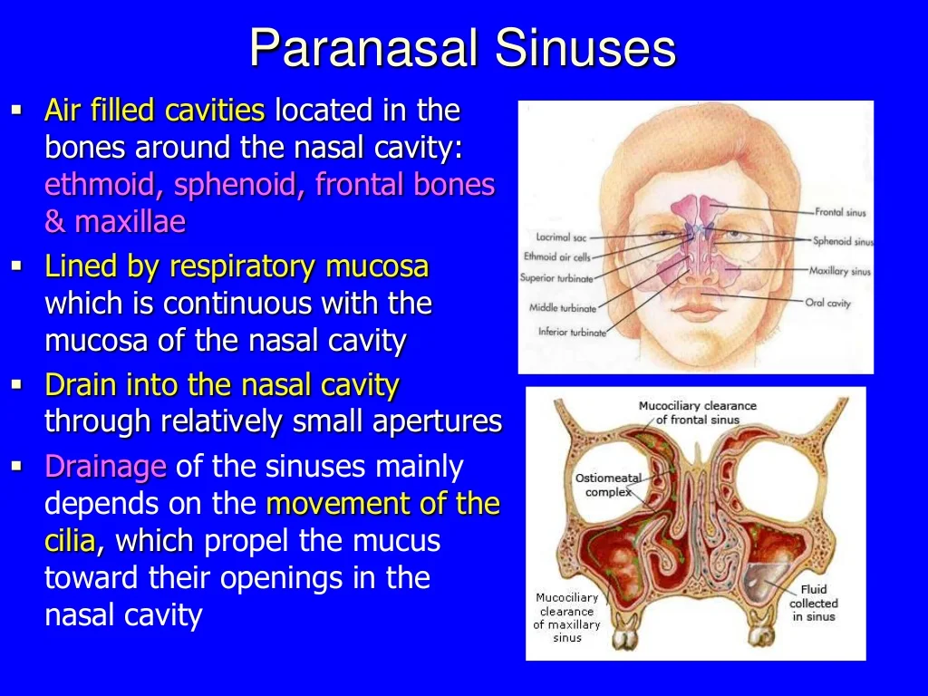
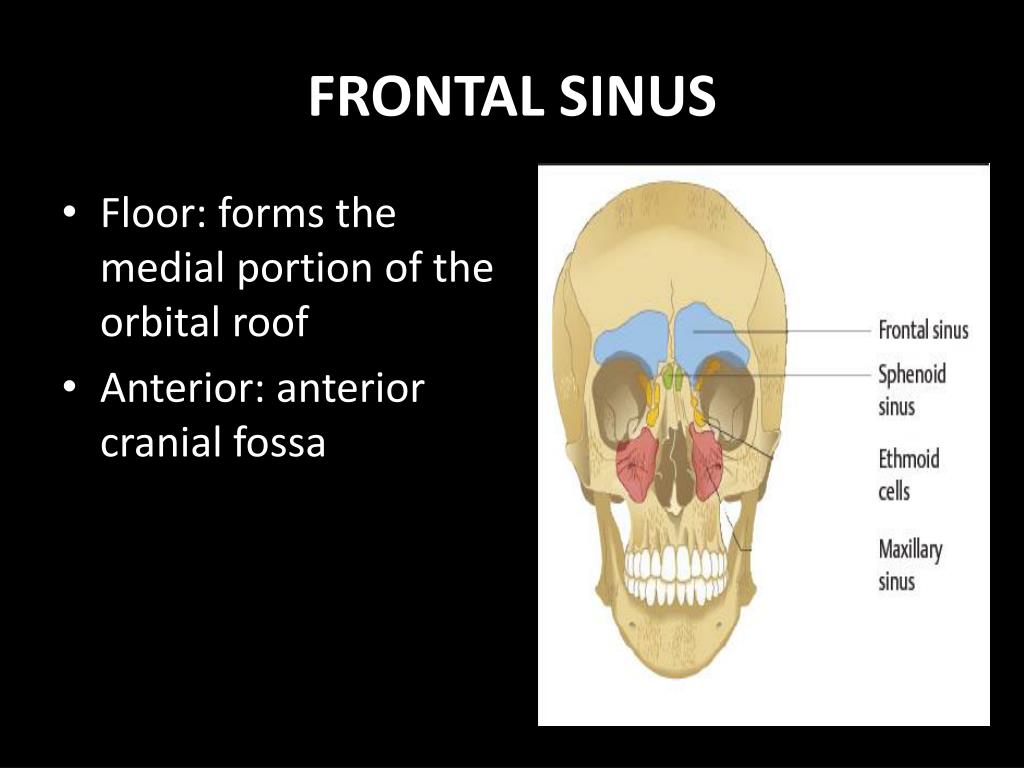
The nose & Paranasal sinuses. The nose & Paranasal sinuses. Bones around the nasal cavity are hollowed out These cavities are the paranasal sinuses and communicate by small aperture with the cavity Lighten the face Resonance of voice Insulator for incoming cold air Determine the position of the orbital cavities. 208 views • 16 slides
PPT MANDIBLE, SINUSES, TEMPORAL BONE PowerPoint Presentation, free download ID6762618
Slide 1 CT IMAGING OF THE PARANASAL SINUSES - BASIC ANATOMY AND IMAGE INTERPRETATION Benjamin Y. Huang, MD, MPH University of North Carolina OVERVIEW Indications for imaging chronic sinusitis Scan technique Basic anatomy of the paranasal sinuses Anatomic variants which predispose to sinusitis and surgical complications

Paranasal sinuses
ß"‰1¨_þq‡"ÅþÐÓŒO±,, ²ø« r ¢e5p'«³)3ÒÍ瀲ñt¾Òi'_¢ ]ˆ÷ L y‰Í Z *¤ °hÏ2ƒÌxÁ Ž¢ \ýVªbZ`]Po²È ôF%´.°íÝèZ‹dÉM{ƒÆzò è¦XÖÙ„öáš¡¿ùöq - ñ ò-‚ç àaÔë ŽÓ‰ öG°:ij›Ä#ø £îp8LNÃàŽ ®õ€t lK™»_y>SÊì&IÜ~Öˆý^ Û;±à÷ ß èX Â(é÷ Œk?6kØ.
PPT NASAL CAVITY & PARANASAL SINUSES PowerPoint Presentation ID6150524
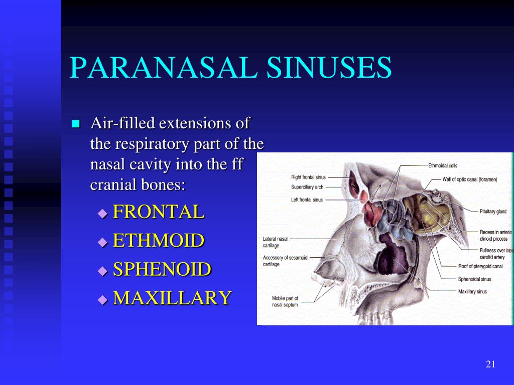
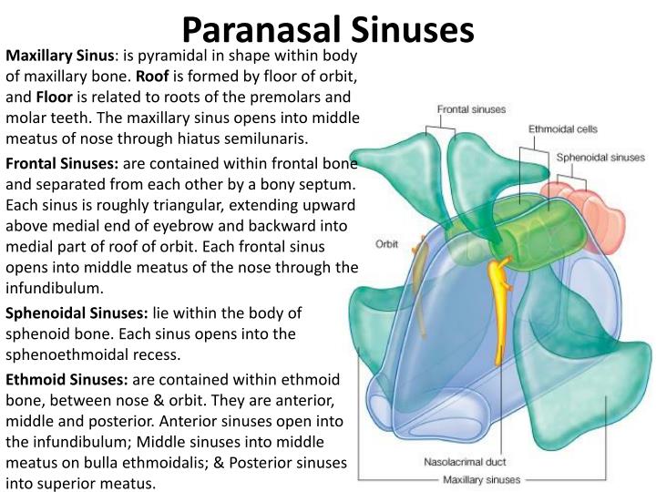
1/4 Synonyms: Antrum of Highmore, Maxillary paranasal sinus , show more. The paranasal sinuses are paired and symmetrical, air-filled cavities situated around the nasal cavity. Paranasal sinuses are found in three bones of the neurocranium (braincase), the frontal bone, ethmoid bone, and sphenoid bone.
Nasal cavity
Vocal resonance • Nasal cavity & paranasal sinus cavity provide vocal resonance for nasal consonants M, N & nG • De-nasal voice is seen in nose block. Nasal consonants M, N & nG are uttered as B, D & G respectively. Nasal reflexes 1. Smell reflex: increasessecretions of saliva & gastric juice 2. Naso-pulmonary reflex:Chronic, severe nasal.

PPT The Axial Skeleton PowerPoint Presentation, free download ID2098222
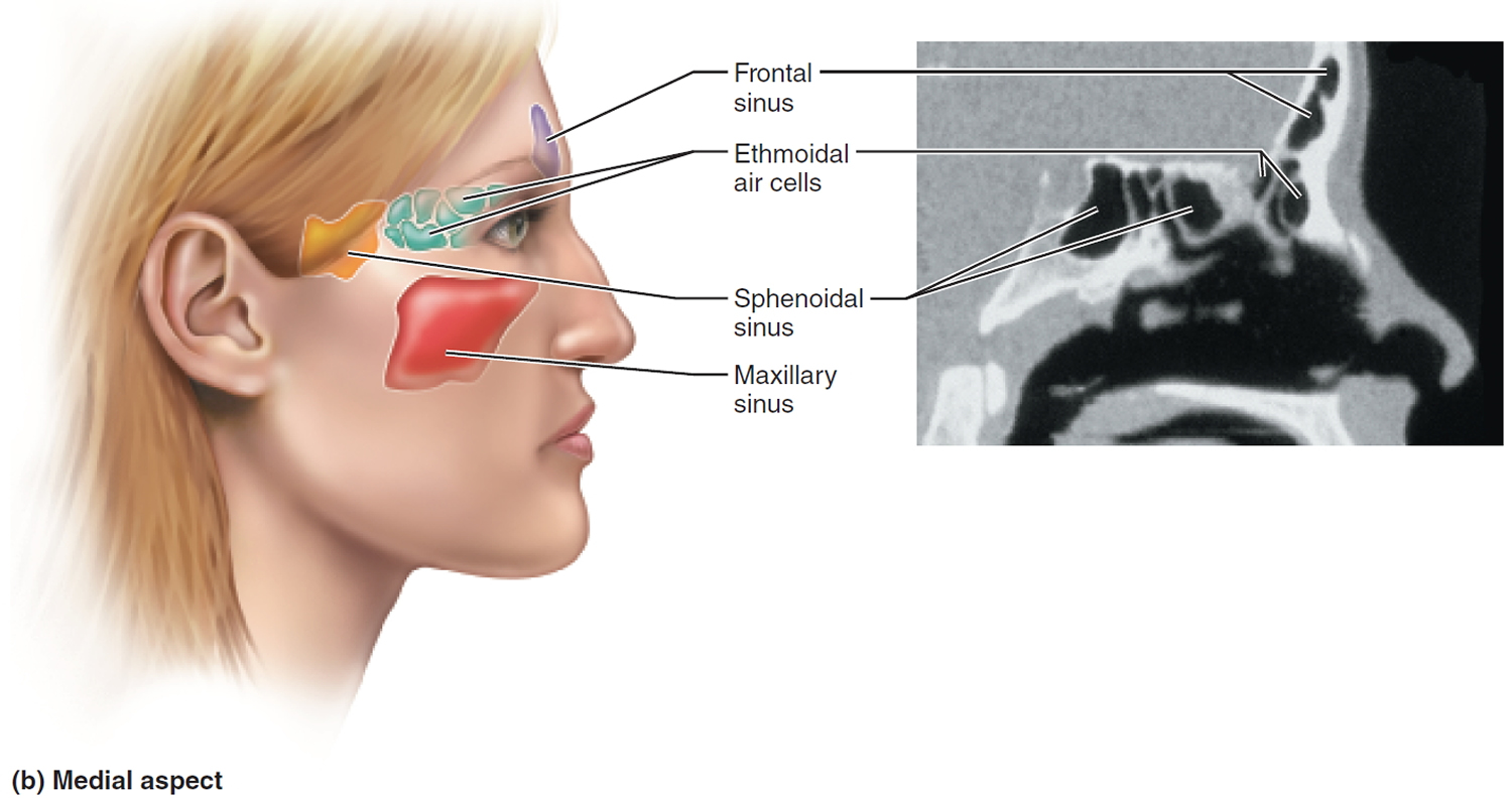
The maxillary sinus is the largest paranasal sinus and lies inferior to the eyes in the maxillary bone. It is the first sinus to develop and is filled with fluid at birth. It grows according to a biphasic pattern, in which the first phase occurs during years 0-3 and the second during years 6-12. The earliest phase of pneumatization is directed.
PPT PARANASAL SINUSES Anatomy, Physiology and Diseases PowerPoint Presentation ID2649604
Paranasal sinuses. Feb 8, 2019 •. 15 likes • 3,514 views. Dr Sudeep Madhusudan Chaudhari Pediatric Dentist.

Image result for paranasal sinuses communication Paranasal sinuses, Sinusitis, Cavities
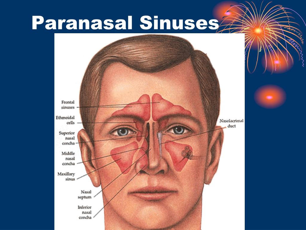
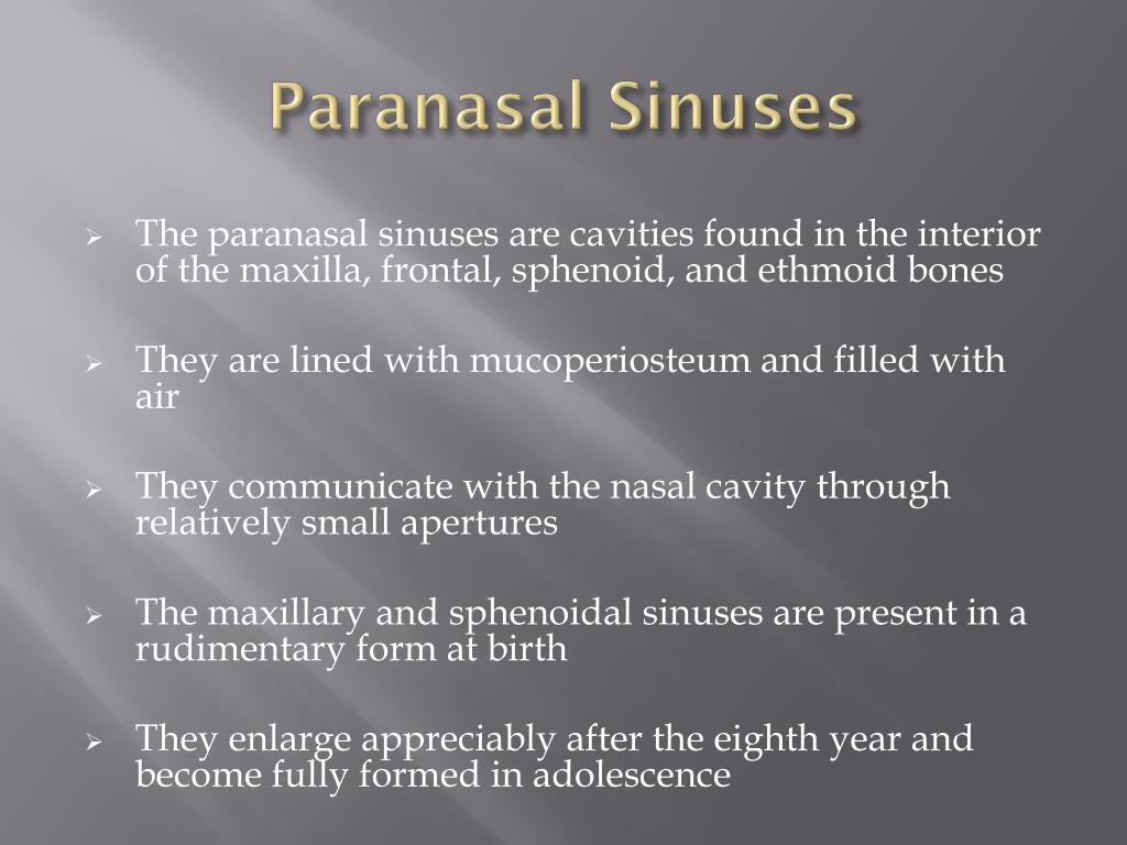
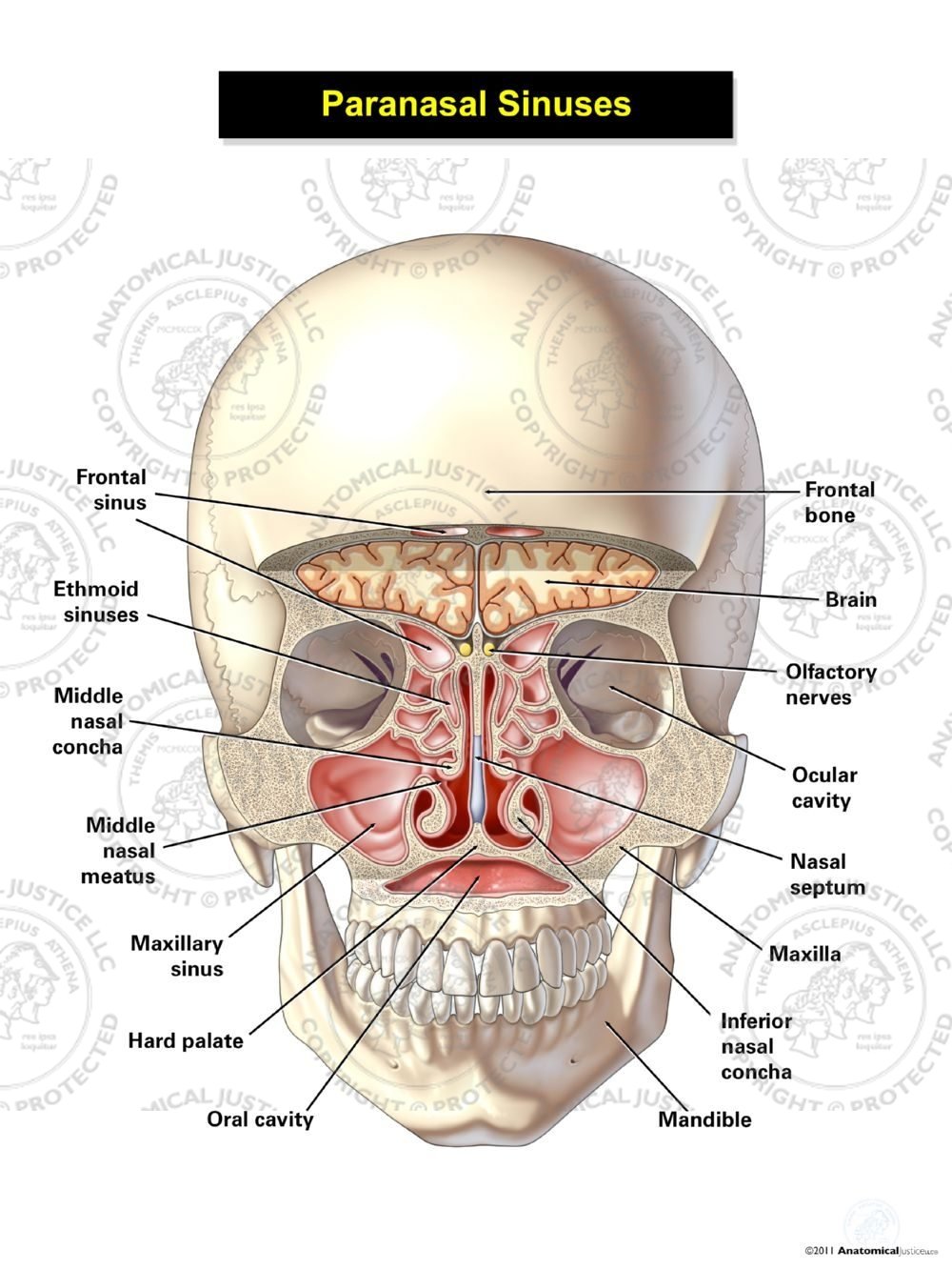
Last updated: April 1, 2021 Revisions: 21 format_list_bulleted Contents add The paranasal sinuses are air-filled extensions of the nasal cavity. There are four paired sinuses - named according to the bone in which they are located - maxillary, frontal, sphenoid and ethmoid.

frontal cut anatomy
1 Surgical Anatomy of the Paranasal Sinus M. PAIS CLEMENTE The paranasal sinus region is one of the most complex areas of the human body and is consequently very diffi-cult to study.
Sinuses Anatomy Of The Head
Anatomy of Paranasal Sinuses | PPT Anatomy of Paranasal Sinuses Jul 6, 2015 • 43 likes • 11,617 views Download Now Download to read offline M Meghna Rai Follow Recommended Anatomy of nose and paranasal sinuses Vinay Bhat 84.4K views • 52 slides Anatomy & development of the middle ear Sayan Banerjee 880 views • 52 slides
Chronic Sinusitis Causes, Symptoms, Surgery, and Treatment
Anatomy of nose and paranasal sinuses Aug 25, 2012 • 336 likes • 84,391 views Download Now Download to read offline Health & Medicine Technology Vinay Bhat Assistant Professor at Pondicherry Institute of Medical Sciences Follow Recommended Anatomy of nose and para nasal sinuses . by DR. MD. KHURSHID PERVEJ. GMC PATIALA kbristi12

Paranasal sinuses
Nose and Sinuses Medical Theme Presentation Free Google Slides theme and PowerPoint template The nose. well, it's very important. It is the one that allows us to smell everything around us. Unfortunately, it is not immune to suffering from diseases.

What Sinuses Drain Into The Middle Meatus Best Drain Photos
Paranasal Sinuses. When there is facial trauma, the paranasal sinuses can act as a "crumple zone" protecting the more delicate structures of the brain from injury. Dural Venous Sinuses. The emissary veins, which traverse the cranium and enter the dural venous sinus system, are a pathway that infection can enter the brain. This phenomenon is.

Paranasal Air Sinuses location, Functions, Relations and Applied
Maxillary Sinus (within the maxillary bones): The largest among all the paranasal sinuses [2], these two conical cavities are located on the two sides of the nose, above the upper teeth, and below the cheeks [4]. Ethmoid Sinus (within the ethmoid bones): Three to eighteen [5] air cells present in the ethmoid labyrinth, on both sides of the nose, between the eyes [6, 7].
PPT Nasal Cavity & Paranasal sinuses PowerPoint Presentation ID1827415
Anatomy, Head and Neck, Nose Paranasal Sinuses - StatPearls - NCBI Bookshelf The uncinate process is a delicate, sickle-shaped, bony part of the ethmoid bone, covered by mucoperiosteum, medial to the ethmoid infundibulum, and lateral to the middle turbinate.
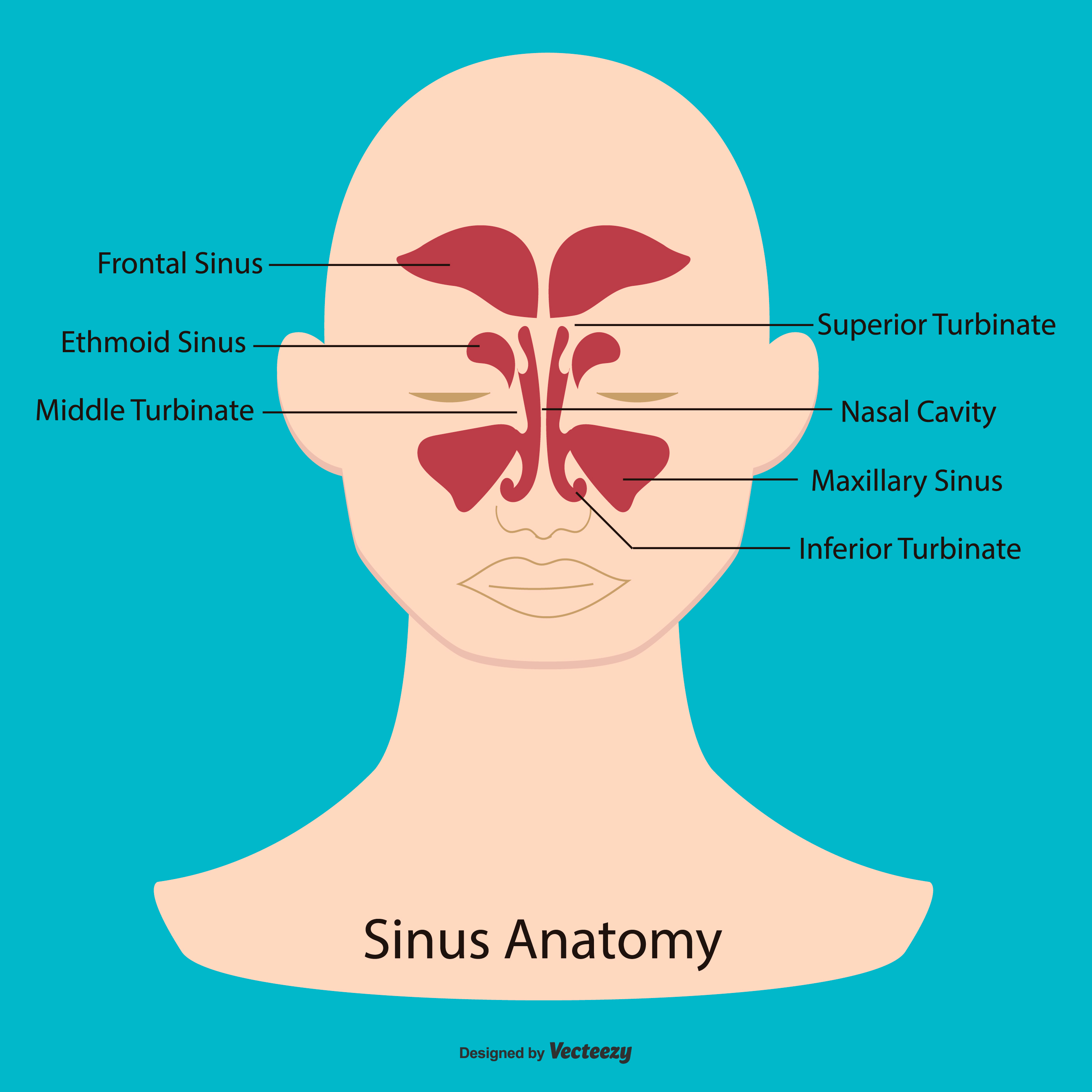
Sinus Anatomy Illustration 172412 Vector Art at Vecteezy
PPT - Paranasal Sinuses PowerPoint Presentation, free download - ID:6525008 Presentation Download 1 / 21 Download Presentation >> Paranasal Sinuses Nov 13, 2014 280 likes | 788 Views Paranasal Sinuses. Kristina Fatima Louise P. Garcia Group 5A1. Embryology of the Paranasal Sinuses.