
Anterior Triangle of the Neck Subdivisions TeachMeAnatomy
Contents Anatomical triangles Anterior triangle Muscular triangle Carotid triangle Submandibular (digastric) triangle Submental triangle Posterior triangle Occipital triangle Supraclavicular (omoclavicular) triangle Clinical significance Jugular venous pressure Carotid artery pulsation Cricothyroidotomy Sources + Show all Anatomical triangles
Anterior Triangle of Neck Anatomy QA
It passes obliquely across the neck, stretching from the sternum and clavicle below to the mastoid process of the temporal bone and superior nuchal line of the occipital bone above. Each major triangle can be further subdivided into even smaller triangles. The triangular space anterior to the sternocleidomastoid is called the anterior neck.
Anterior Triangle of Neck Anatomy QA
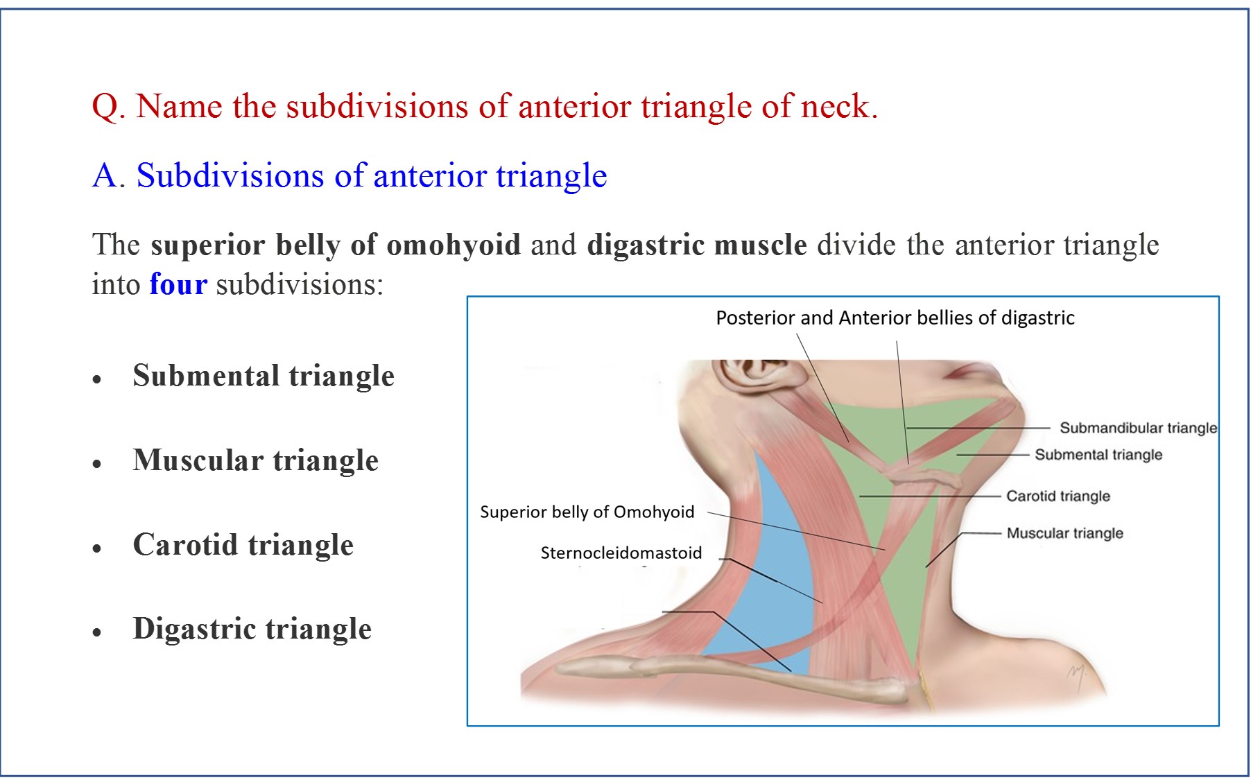
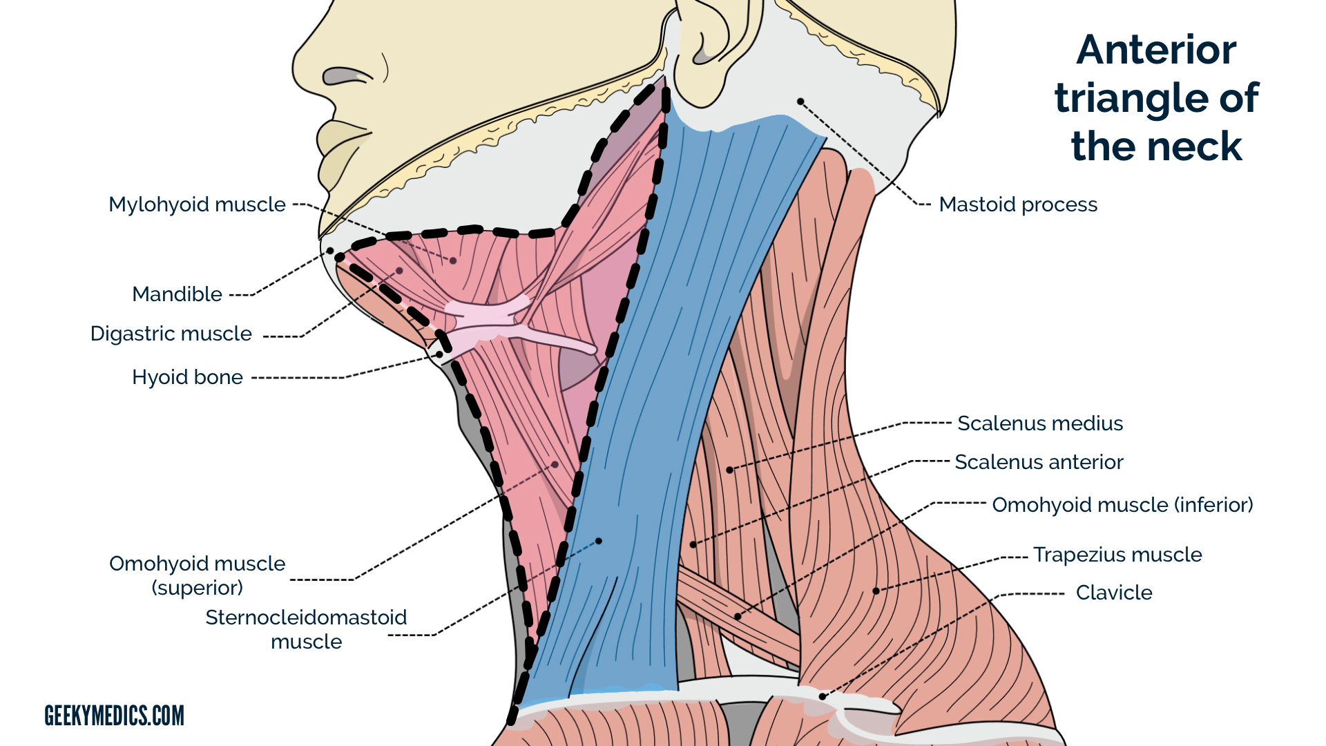
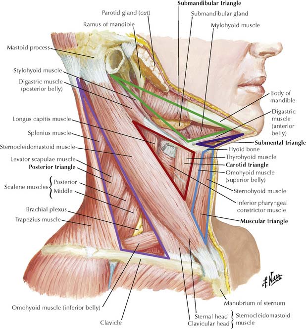
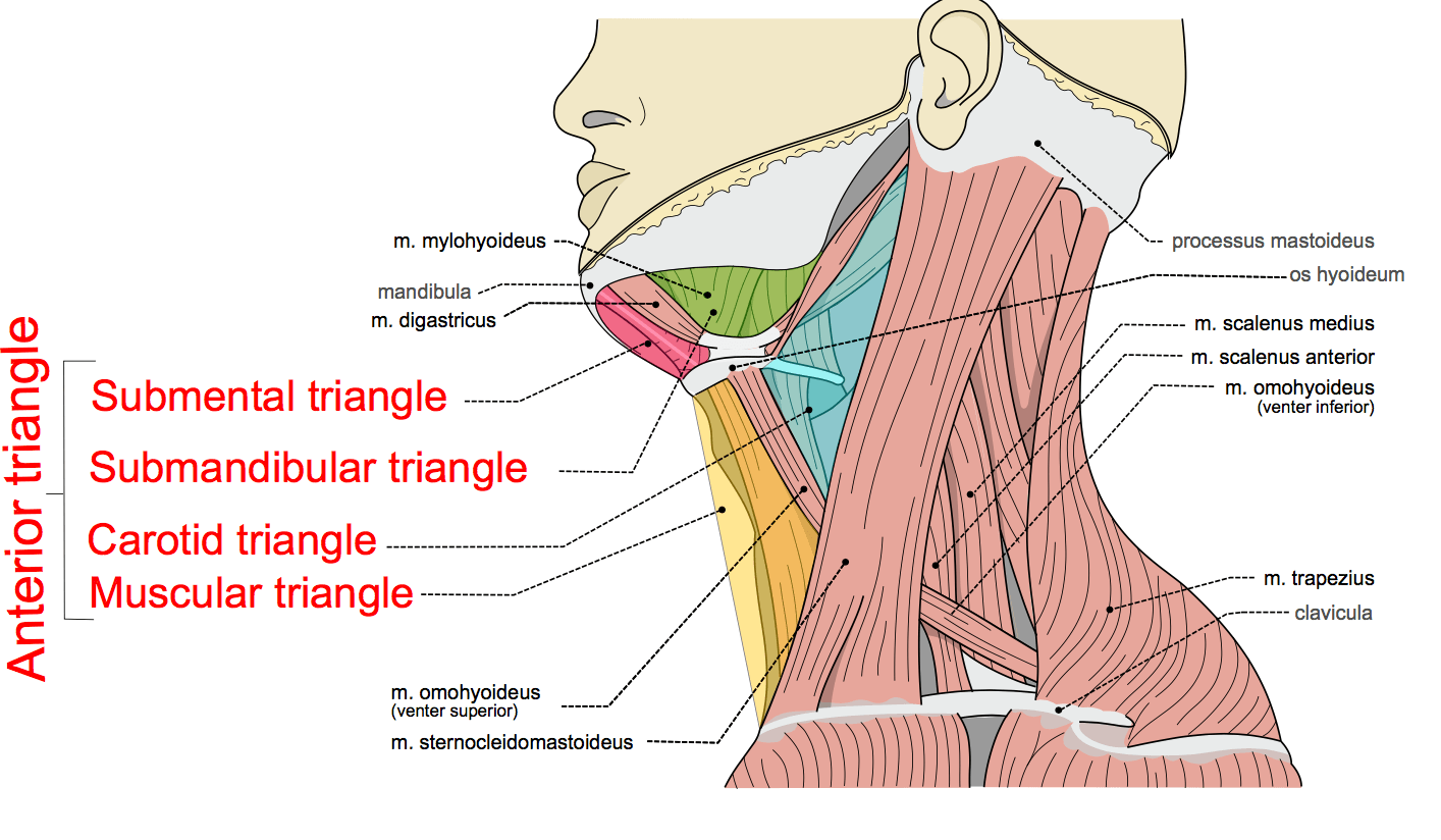
Contain glands, nerves, vessels, and lymph nodes Sternocleidomastoid muscle (SCM) divides the neck into the 2 major neck triangles: Anterior triangle: subdivided into smaller triangles Muscular triangle Carotid triangle Submandibular triangle Submental triangle

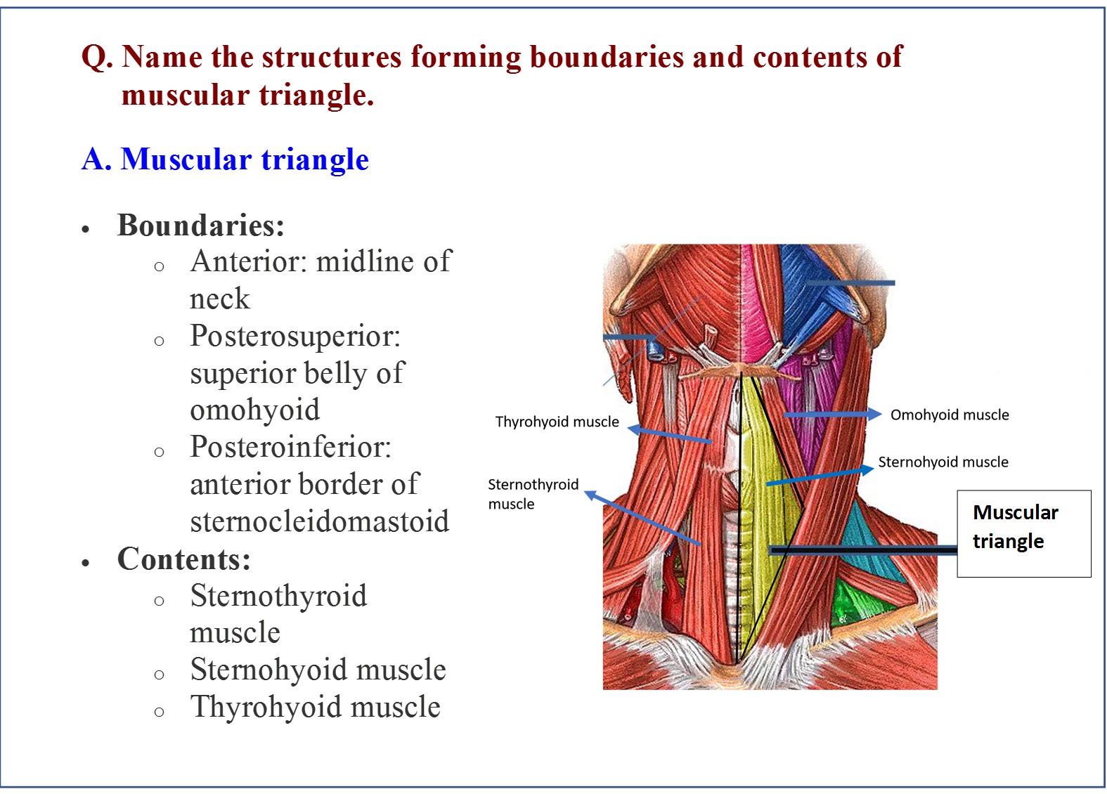
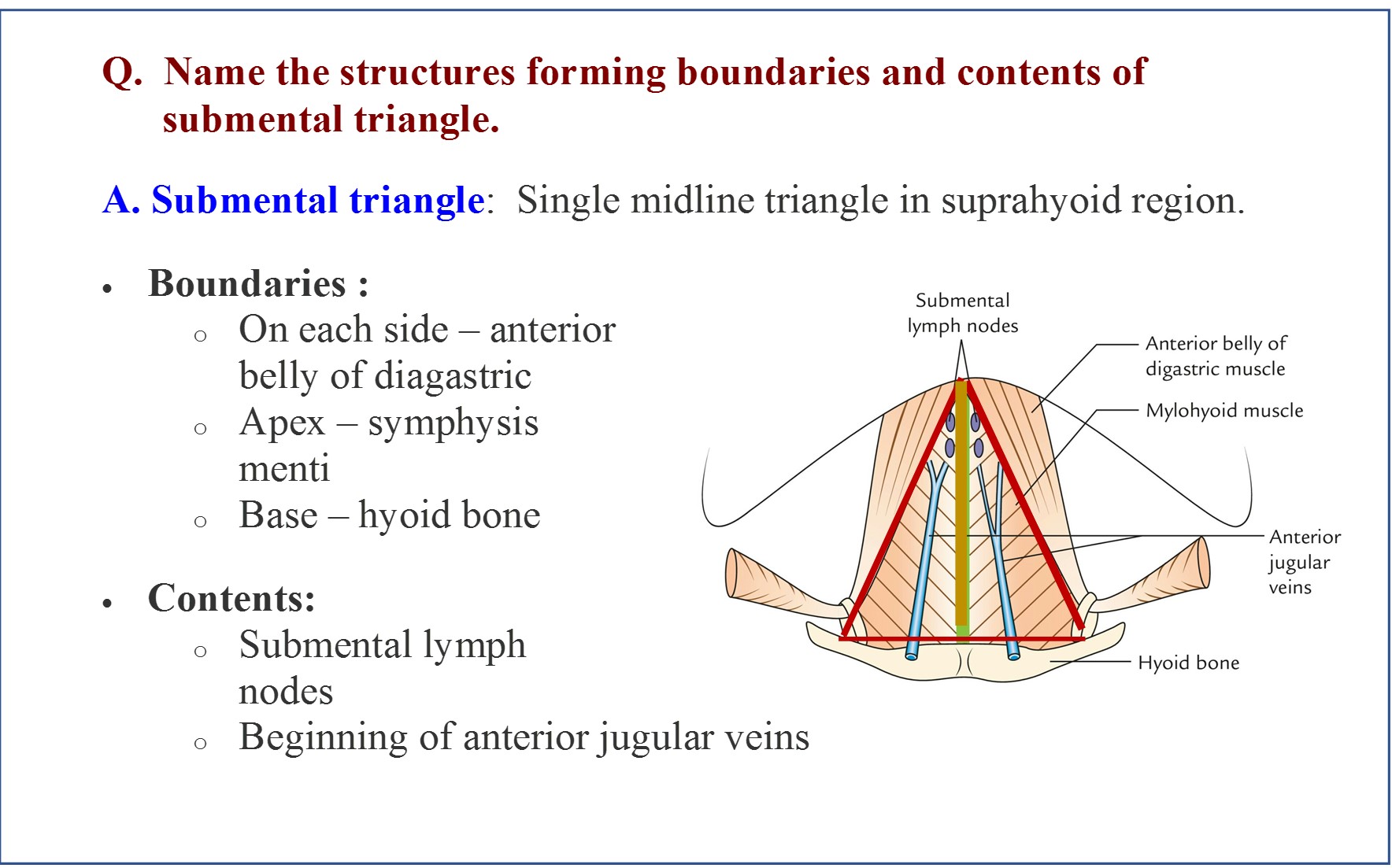
Anterior Triangle of Neck Submental and Muscular triangles Boundaries and Contents
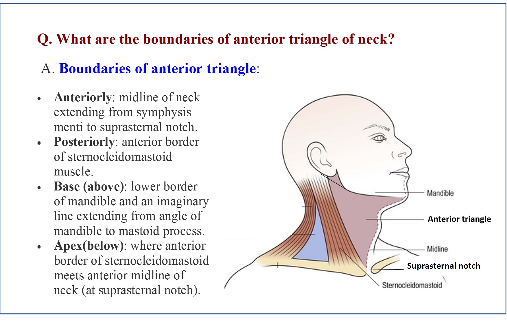
The anterior triangle is a region located at the front of the neck. In this article, we shall look at the anatomy of the anterior triangle of the neck - its borders, contents and subdivisions. Note: it is important to note that all triangles mentioned here are paired; they are located on both the left and the right sides of the neck. Borders
Anterior Triangle of Neck Anatomy QA
The anterior triangle is roofed by the investing layer of deep cervical fascia overlying which is the platysma muscle and subcutaneous fat. The platysma muscle crosses the lower border of the mandible to become continuous with some of the muscles of facial expression. Inferiorly it ends by blending with the thin connective tissue overlying the.
Anterior Triangle of the Neck Earth's Lab
The anterior cervical region or triangle can be topographically located at the anterior portion of the neck. It spans seven levels of cervical vertebrae (C1-7). The anterior triangle is a region bounded superiorly by the inferior border of the mandible, laterally by the anterior median of the sternocleidomastoid muscle, and inferiorly by the jugular and clavicular notch of the manubrium.

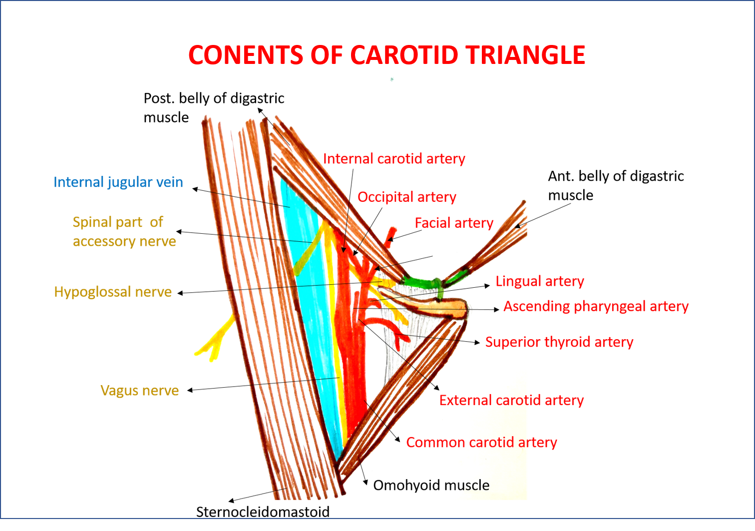
Carotid triangle of neck/ Anatomy /Simplified Boundaries & contents/Anterior triangle of neck
The neck is divided in two major triangles: anterior and posterior, based mainly on the borders of the sternocleidomastoid, or SCM, and trapezius muscles, as well as other muscular and bony structures found in the neck. These regions provide a clear location regarding the structures, injuries or pathologies involving the neck.
Neck Lump Examination OSCE Guide Geeky Medics
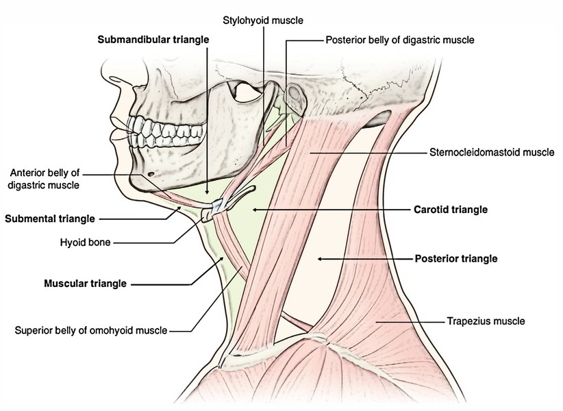
Watch on The sternocleidomastoid muscle obliquely crosses the neck to form the division between the two major neck triangles: anterior triangle and posterior triangle. Both triangles are further divided into sub-triangles. [2] [3] Anterior Triangle Digastric/Submandibular Triangle Carotid Triangle Muscular Triangle Submental Triangle
Triangles of the neck Anatomy, borders and contents Kenhub
#anatomy #head #neckDonation Link: https://paypal.me/studentlamedicina?locale.x=en_UShttps://www.instagram.com/anatomy.knowledge/The anterior triangle of th.
Anterior Triangle of Neck Anatomy QA
The content of the neck is grouped into 4 neck spaces, called the compartments. Vertebral compartment: contains cervical vertebrae and postural muscles.. triangles of the neck. The anterior triangle of the neck is made by the anterior border of the sternocleidomastoid muscle, the inferior border of the mandible and the midline of the neck.
neck triangles
Contents The anterior triangle is subdivided into three paired triangles and a single midline triangle: Paired triangles: digastric triangle muscular triangle carotid triangle Single midline triangle: submental triangle Boundaries anterior: median line of the neck posterior: anterior margin of sternocleidomastoid muscle
The Neck Basicmedical Key
Book Contents Navigation. Contents. Introduction. I. Musculoskeletal Anatomy presented in MJBS. Spine, Spinal cord, Spinal nerves, and Back. Posterior Triangle of the Neck.. Anterior triangle of the Neck. IV. Abdominal Anatomy presented in HNG. Anterior Abdominal Wall, Inguinal Region, and Descent of the Testis.

Anterior Triangle of Neck Submental and Muscular Triangles Anatomy QA
The anterior triangles refer to bilateral anatomic subdivisions of the neck comprising the anterior surface of the neck, deep to the superficial cervical fascia and platysma muscle. Laterally, the anterior triangle is bounded by the anterior border of the sternocleidomastoid muscle. Its superior border is the inferior border of the mandible.
Triangles of the Neck Part 1 The Anterior Triangle Medical Exam Prep
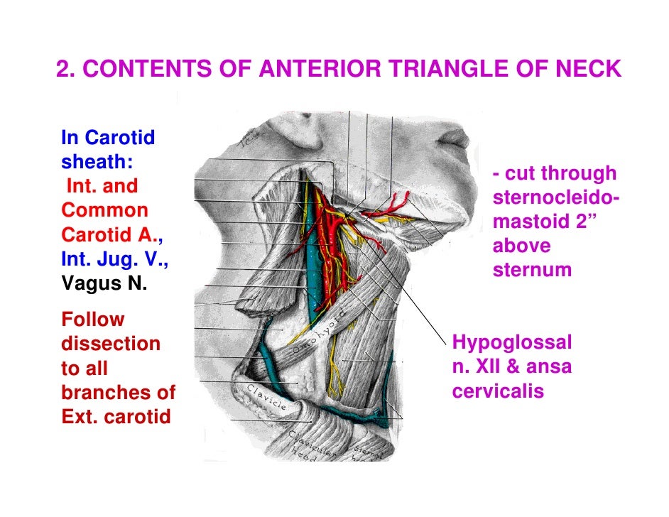
The anterior triangle is formed by the inferior border of the mandible, the anterior border of sternocleidomastoid and the sagittal plane in the midline of the neck. It has 4 main subdivisions: The carotid triangle marks the position of the bifurcation of the common carotid artery, the internal jugular vein and cranial nerves X & XII.
Anterior Triangle of Neck Anatomy QA
The anterior cervical triangle is bounded by the midline of the neck, the anterior border of the sternocleidomastoid muscle (SCM), and the inferior border of the mandible [ 3 ]. This triangle is typically subdivided into three paired and one unpaired triangle.
Anterior Triangle of Neck Anatomy QA
From a surgical perspective, the neck is usually divided into two triangles, namely, the "anterior triangle," which consists of three-and-a-half triangles, and the "posterior triangle," which consists of two triangles (Fig. 1.1).The sternocleidomastoid (SCM) muscle is the key to understanding both of these triangles [].The neck also contains triangles such as the suboccipital triangle.